Harriet Hosmer and the Anatomy of Neoclassical Sculpture
by Elizabeth KetoRecipient of the 2025 Mervat Zahid Cultural Foundation Prize, of the Dahesh Museum of Art
Elizabeth Keto is a PhD candidate in the history of art at Yale University. Her dissertation, “Reconstruction’s Objects: Art in the United States South, 1865–1900,” explores the role played by images, objects, and their makers in the reshaping of social and political order in the decades following emancipation and the end of the Civil War. Her research has been supported by the Smithsonian American Art Museum and the National Museum of American History. She is the 2025–27 Twenty-Four-Month Chester Dale Fellow at the Center for Advanced Study in the Visual Arts at the National Gallery of Art, Washington, DC.
Email the author: elizabeth.keto[at]yale.edu
Citation: Elizabeth Keto, “Harriet Hosmer and the Anatomy of Neoclassical Sculpture,” Nineteenth-Century Art Worldwide 25, no. 1 (Spring 2026), https://doi.org/10.29411/ncaw.2026.25.1.3.
This work is licensed under a Creative Commons Attribution-NonCommercial 4.0 International License  unless otherwise noted.
Your browser will either open the file, download it to a folder, or display a dialog with options.
In 1850, Harriet Goodhue Hosmer (1830–1908) made three ink drawings on paper. Created when the artist was twenty years old, each study measures about two by three feet and is executed in lines and washes of grey and black (figs. 1, 2, 3). Each depicts a human figure, stripped of skin to display the ropes and ribbons of tissue beneath, with hundreds of muscles labeled in minute Latin script. Two of the studies show a similarly posed male figure viewed from the front (see fig. 1) and the back (see fig. 2): The figure’s hips are slung in gentle contrapposto, the index finger of its proper left hand points downward, and the right hand gestures with an open palm. The other study (see fig. 3) depicts a male figure from the side: The figure’s proper right knee is bent as if to mount an invisible stair, the right arm is raised in a gesture of salutation or expostulation, and the knuckles of his left hand rest lightly on a boxy surface. Depicted without skin—a convention of anatomical representation known as écorché, after the French word for “flayed”—Hosmer’s figures are animated yet not alive, suspended between human and thing, artistic subject and scientific object.
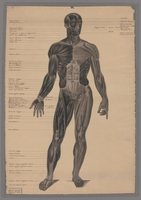
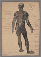
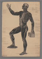
Each drawing is signed and dated at the bottom left. Hosmer’s proud declaration in black ink, “HHosmer pinxit,” or “H[arriet] Hosmer painted [it],” suggests that she viewed the drawings not merely as academic exercises but also as works of art. As such, these anatomical studies constitute the earliest extant objects in Hosmer’s oeuvre. All three are now archived among the artist’s papers at the Schlesinger Library of the Radcliffe Institute for Advanced Study at Harvard University. The material condition of the studies offers additional evidence of their importance to the sculptor. Clusters of tiny pin holes at each corner of the drawings corroborate a description of Hosmer’s studio published in 1852, which noted that “immense anatomical plates, executed by the lady herself, adorned the walls.”[1] She displayed the studies alongside the other scientific and medical objects—including, reportedly, a complete human skeleton—that littered her workspace at her family home in Watertown, Massachusetts.[2] Later in 1852, Hosmer traveled to Rome to begin her professional career as a neoclassical sculptor, bringing with her several proofs of her skill: a daguerreotype of her first bust in marble, a certificate from her course of study in anatomy, and her anatomical drawings.[3] Yet despite their apparent importance to the artist, these works on paper have not yet been the subject of art historical analysis, nor have the scientific interests and education which produced them been given critical attention.[4]
Hosmer’s anatomical drawings reflect the convergence of neoclassical aesthetics and medical science in the depiction of the human body. Medical education, surgical and anatomical images, and the practice of dissection provided the basis for these drawings and supported Hosmer’s professional formation more broadly. I argue that Hosmer’s anatomical training and the patronage which enabled it linked the artist to institutions and discourses that mediated notions of racial and gender normativity, difference, and hierarchy in the nineteenth-century United States.
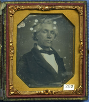
Hosmer became one of the nineteenth century’s most celebrated neoclassical sculptors and one of its best-known women artists in any medium. The three drawings testify to her necessary preparation for the practice of figurative sculpture by the study of human anatomy. In the winter of 1850 and spring of 1851, Hosmer studied under the tutelage of Joseph Nash McDowell (1805–68), a surgeon and professor of anatomy in the medical department of the University of Missouri in St. Louis (fig. 4).[5] Though anatomy was a standard element of nineteenth-century artistic education, Hosmer’s decision to pursue the subject at a medical school, rather than an art academy, meant that her experience differed in significant ways from that of many academically trained painters and sculptors who would become her peers in Rome.[6] While dissection was the basis of anatomical instruction in medical schools, most European art academies “did not incorporate dissecting rooms into their buildings,” given public distaste for the practice and the danger of infection from contact with dead bodies prior to the development of antiseptic practices and the discovery of formaldehyde.[7] Students in the academies instead learned to depict the human body by studying plaster casts of corpses with the skin removed to show musculature, known as écorchés; casts of antique statues; and live nude models.[8] But with few art academies established in the mid-nineteenth-century United States comparable to those of Paris or London, some of the country’s aspiring painters and sculptors, like Hosmer, turned to medical schools to procure anatomical knowledge by studying dissected bodies alongside physicians in training.[9]
Hosmer’s position as a woman artist rendered her training in anatomy more remarkable, a fact which she cannily used in her self-mythologizing. Decades after her sojourn in St. Louis, Hosmer recalled her reasons for traveling west to the city’s medical college in an interview with the St. Louis Globe-Democrat in 1888: “In those days it was another thing for our sex to enter a medical college . . . Eastern schools were positively closed.”[10] As historian Kate Culkin has noted in her critical biography of Hosmer, the artist’s gendered exclusion from medical education in the Northeast is a standard element of Hosmer’s life story, repeated in numerous later biographies and other accounts.[11] But as a matter of historical fact, it is not entirely true. Hosmer most likely could have accessed anatomical training at medical institutions closer to her family’s home in Watertown. The Boston Female Medical College, first of its kind in the nation, was established in 1848, while Elizabeth Blackwell had recently become the first woman to attend medical school in the United States, after her acceptance at Geneva Medical College in New York in 1847.[12] The artist’s father, Hiram Hosmer, was himself a doctor; he instructed Harriet’s cousin Alfred in anatomy and could surely have taught his daughter as well.[13]
The persistence of this narrative of exclusion and exceptionalism may be due not only to Hosmer’s self-construction but also to the importance of what Linda Nochlin famously termed “The Question of the Nude” in feminist art history.[14] In her article of 1971, “Why Have There Been No Great Women Artists?,” Nochlin asserted that “the complete unavailability to the aspiring woman artist of any nude models at all, male or female” during the eighteenth century and most of the nineteenth effectively barred women from classical subjects and confined them to less prestigious genres of representation.[15] Art historians have critically nuanced Nochlin’s position in recent years, but the question of whether, how, and under what conditions nineteenth-century women artists could study naked human bodies remains linked to broader analytical problems and historiographical debates.[16] Hosmer’s ability to negotiate access to anatomical instruction makes her a case study in what historian and art historian Paris A. Spies-Gans terms the “tangled patterns of exclusion, inclusion, and assimilation” that contoured the relationships between women artists and the institutions of artistic education and professionalization in the nineteenth century.[17]
The aspiring sculptor’s decision to pursue her anatomical studies in St. Louis likely reflected a convergence of personal and professional inducements. Hosmer had attended school in Lenox, Massachusetts, from 1847 to 1849.[18] There, she began an intimate and lifelong friendship with Cornelia Crow, later Cornelia Crow Carr, daughter of the wealthy St. Louis dry-goods merchant Wayman Crow.[19] The opportunity to live for months in the same household as Cornelia was doubtless appealing, as was the opportunity to cultivate the favor of Cornelia’s father, who would become the most important patron of Hosmer’s career. Hosmer may also have wished to study with McDowell because the anatomist had previous experience instructing sculptors, including the celebrated Hiram Powers (1805–73).[20] Crow, a civic leader, possessed whatever influence might have been necessary to persuade McDowell to accept Hosmer as a student.
Hosmer’s three anatomical drawings constitute an interpretively complex record of her time in St. Louis under McDowell’s tutelage. In this article, I take seriously Hosmer’s declaration that her anatomical drawings were works of art and examine the role they played in her artistic formation. These drawings reflect not only her absorption of medical knowledge but also her close engagement with the visual and epistemological relationship between art and anatomy in the mid-nineteenth century. Her studies draw upon an influential set of French anatomical illustrations dating to the 1830s, which included novel strategies of medical and surgical depiction. Yet the three figures from that larger publication which Hosmer selected to reproduce reflect instead a much older tradition of artistic anatomy.[21] Hosmer depicted écorché figures modeled in elegant contrapposto, their gestures echoing illustrations that had first appeared in sixteenth-century anatomical texts. Like their prototypes in the work of Andreas Vesalius (1514–64) and other early modern anatomists, Hosmer’s figures synthesize scientific knowledge and artistic imagination, combining the internal bodily structures revealed by anatomy with the poses and proportions of classical sculpture.
Hosmer’s anatomical training was a professional necessity, but it also influenced the ways in which both the artist and her critics understood her early works in marble. Her sculptures became identified with a medicalized aesthetics linked to the construction of gender, sexuality, and race. Existing scholarly literature on Hosmer has dealt extensively with issues of gender and sexuality, but it has contended less often with the question of race in her work.[22] Yet Hosmer’s education in St. Louis bound her professional formation to the discourses of racial science, the institution of chattel slavery, and the profits of racial capitalism. Without a careful consideration of how the significance of gender and sexuality was articulated and contested through race—and vice versa—in the nineteenth-century United States, we cannot attend to the complexity of Hosmer’s politics, nor that of her sculptural practice.[23]
Drawing on Anatomy
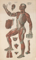
Hosmer’s drawings themselves offer important evidence of the medical texts, images, and practices to which she was introduced during her training in St. Louis. Although she signed the three anatomical studies as her own, the figures are in fact hand-drawn reproductions, closely based upon plates 3, 5, and 7 of the Anatomie élémentaire en 20 planches (Elementary Anatomy, in Twenty Plates), an elephant folio set of exquisitely detailed lithographs after drawings by the French artist Nicolas Henri Jacob (1781–1871), in collaboration with the anatomist Jean-Marc Bourgery (1797–1849) (figs. 5, 6, 7).[24] Published in Paris between 1836 and 1839, the large-scale lithographs supplemented Bourgery and Jacob’s magisterial anatomical atlas and surgical manual, the Traité complet de l’anatomie de l’homme (Complete treatise of the anatomy of man), which appeared in eight illustrated volumes between 1831 and 1854.
Fig. 1. Harriet Goodhue Hosmer, Anatomical Drawing, 1850. Pen, ink, and wash on paper. Papers of Harriet Goodhue Hosmer, Schlesinger Library, Radcliffe Institute for Advanced Study, Harvard University, Cambridge. Artwork in the public domain; image courtesy of the Schlesinger Library, Radcliffe Institute, Harvard University.
Fig. 5. After Nicolas Henri Jacob, Myologie et aponeurologie, plan anterieur (Myology and Aponeurology, Anterior View), 1836/1839. Lithograph. Published in Jean-Marc Bourgery and Nicolas Henri Jacob, Anatomie élémentaire en 20 planches (Paris, 1836/1839), n.p., plate 3. Artwork in the public domain; image courtesy of the Internet Archive.
Fig. 2. Harriet Goodhue Hosmer, Anatomical Drawing, 1850. Pen, ink, and wash on paper. Papers of Harriet Goodhue Hosmer, Schlesinger Library, Radcliffe Institute for Advanced Study, Harvard University, Cambridge. Artwork in the public domain; image courtesy of the Schlesinger Library, Radcliffe Institute, Harvard University.
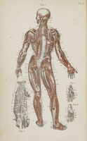
Fig. 6. After Nicolas Henri Jacob, Myologie [et] aponéurologie, plan postérieur (Myology [and] Aponeurology, Posterior View), 1836/1839. Lithograph. Published in Jean-Marc Bourgery and Nicolas Henri Jacob, Anatomie élémentaire en 20 planches (Paris, 1836/1839), n.p., plate 5. Artwork in the public domain; image courtesy of the Internet Archive.
Fig. 3. Harriet Goodhue Hosmer, Anatomical Drawing, 1850. Pen, ink, and wash on paper. Papers of Harriet Goodhue Hosmer, Schlesinger Library, Radcliffe Institute for Advanced Study, Harvard University, Cambridge. Artwork in the public domain; image courtesy of the Schlesinger Library, Radcliffe Institute, Harvard University.
Fig. 7. After Nicolas Henri Jacob, Myologie [et] aponéurologie, plan lateral (Myology [and] Aponeurology, Side View), 1836/39. Published in Jean-Marc Bourgery and Nicolas Henri Jacob, Anatomie élémentaire en 20 planches (Paris, 1836/1839), n.p., plate 7. Artwork in the public domain; image courtesy of the Internet Archive.
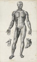
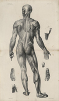
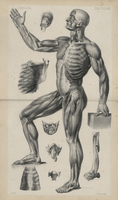
These three plates in Bourgery and Jacob’s folio, and Hosmer’s drawings after them, depict the human body rendered without skin to reveal the muscles of the torso, arms, hands, legs, and feet, with individual bands of tissue delineated in feathery grey and black. Each displays several distinct layers of muscular structure. In the front-and-back studies of the figure in contrapposto, the body is divided along a center axis. One half shows, in lighter grey, the aponeuroses, or tissue coverings, of the muscles; the other peers a layer deeper into the body to trace the muscles themselves, in darker tints. The figure shown with bent knee and raised leg is likewise split, with only one half of its torso displaying the rib cage and intercostal muscles. Like their prototypes in Bourgery and Jacob’s illustrations, Hosmer’s figures include no visible genitalia. Indeed, Hosmer’s reproductions are strikingly precise, duplicating even the extraneous, nonscientific details of Bourgery and Jacob’s plates. Her figure in anterior view (see fig. 1), for instance, includes the same low horizon line and cast shadows that appear in the lithograph (see fig. 5). However, Hosmer chose to eliminate the studies of individual muscles and body parts that surround and supplement Bourgery and Jacob’s écorchés, focusing her drawings upon the central figures alone.
Considered as primary sources in the history of medicine, Hosmer’s drawings evidence the transatlantic reception of Bourgery and Jacob’s anatomical images, a reception which likely reflects the influence of French practice in nineteenth-century medical education in the United States. The French clinical tradition emphasized dissection, empiricism, and practical experience, and it became a dominant pedagogical model in the United States during the 1840s.[25] Though antebellum Missouri did not boast the elite medical schools of the Northeast, McDowell was a well-connected and educated man, editor of a medical journal, and likely abreast of new publications and developments in his field. Nephew of a famous surgeon of the early republic, McDowell had studied under Daniel Drake, one of the Midwest’s most eminent physicians, and had himself previously taught at Jefferson Medical College in Philadelphia.[26] It is not impossible that the sumptuous elephant-folio lithographs of the Anatomie élémentaire were among the collections of McDowell’s medical department, and the exactness of Hosmer’s copies suggests that she had access to the original, large-scale plates. Yet the first complete English translation of the Anatomie élémentaire did not appear until 1867, with lithographic plates produced by the Hartford, Connecticut, publishing firm of Kellogg & Bulkeley. As a notice of the new translation in the Boston Medical and Surgical Journal declared in November 1867: “No anatomical plates have ever been published so complete in beauty and accuracy as those of Bourgery and Jacob. Their great cost, however, has kept them out of the libraries of all but the most wealthy of our profession.”[27] It is also possible that Bourgery and Jacob’s images found their way to St. Louis through their piecemeal reproduction in earlier publications. Some of the French lithographs were copied and circulated in the United States from at least the 1850s onward, in texts devoted to both academic medicine and popular science. Among them were several works by the US physician and lecturer Thomas Scott Lambert, whose textbook Popular Anatomy and Physiology (1850) (fig. 8) and set of large folios, Lambert’s Pictorial Anatomy (1851) (fig. 9), reproduced two of the same Bourgery and Jacob figures that were drawn by Hosmer.
Hosmer’s drawings reflect copying from illustrations rather than studying from life, but such copying probably does not reflect the sum of her anatomical practice or knowledge. Like the anatomy courses offered at most other US medical schools in 1850, McDowell’s instruction centrally emphasized the dissection of human cadavers.[28] He held private viewings of dissected cadavers for Hosmer, and she attended his anatomy lectures with the broader student body, which, excepting herself and a single chemistry pupil, was entirely male.[29] Though the professor had reportedly “supposed at first that [Hosmer] was jesting” when she approached him about anatomy lessons, he agreed to teach her once convinced of the “earnest[ness]” of her request.[30] It seems probable that Hosmer’s private studies with McDowell substituted for actually dissecting cadavers herself, which the medical students were required to undertake.
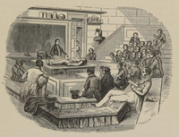
Hosmer walked each day from the Crow house to McDowell’s medical college, which included an anatomy theater, dissecting room, and museum of natural history specimens.[31] An illustration from the title page of an anatomical atlas published in Philadelphia in 1844 gives a sense of how the anatomy theater in the college might have appeared (fig. 10). A few dozen frock-coated young men watch attentively from the benches of the amphitheater while their instructor gestures at the corpse lying ready upon the dissecting table. The vignette suggests the gendered boundaries that usually closed such a space to women and made Hosmer a visible exception to its rules. All the figures in the image are male, save a solitary woman—clad in an apron, perhaps a domestic servant—who appears in the doorway behind the lecturer, her body half-hidden as she peers within. Though entering the typically male-dominated, often profane and bawdy, space of the anatomy theater must have been occasionally uncomfortable, Hosmer retained warm memories of her studies there.[32] In gratitude for McDowell’s tutelage, she carved a bas-relief portrait medallion in marble of the doctor (now lost) and presented it to him in 1852.[33] She wrote nostalgically to Wayman Crow soon after her move to Rome: “How is Dr Mc’Dowell [sic]? Please give him my very best love & tell him I still hope to sit in his Amphitheatre again & hear him lecture.”[34] A few years later, she enclosed a letter to McDowell himself in one of her missives to Crow, adding that McDowell “did me an invaluable service in St. Louis in the Anatomy department & I don’t forget it.”[35]
Another reason to conjecture that the drawings reflect only part of Hosmer’s exposure to medical practices is that large-scale anatomical plates, like those copied by Hosmer, were employed in medical schools as supplements to dissection rather than as substitutes for it. Such images served as both interpretive tools in the presence of actual cadavers and necessary but inferior aides-mémoire in their absence.[36] As the preface to the anatomical atlas of 1844 noted: “Separated from the centre of instruction, and deprived of the advantages of the Dissecting Room, the ideas once so thoroughly acquired begin to fade, and the images once so distinct, become confused and mixed. A recourse to plates, in the absence of dead bodies, is then the only means of refreshing our knowledge.”[37] For Hosmer, the three large drawings she had copied from anatomical plates were likely intended to offer such “recourse” once she departed St. Louis. Traveling home to Massachusetts and determined to retain the knowledge she had gained over months of study, she wrote to Cornelia Crow from a southbound steamboat on the Mississippi River that she had “finished [her] myology and [was] going through it again,” while she also spent “an hour in [her] state room reviewing [her] muscles” every day.[38]
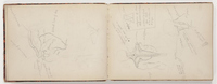
Perhaps because of their origins in medical education, Hosmer’s anatomical drawings are visibly distinct from the anatomical studies produced by many of her artistic contemporaries. Her slightly younger compatriot John La Farge (1835–1910), trained in a Parisian atelier, filled a sketchbook with anatomical drawings in 1864 (fig. 11). La Farge’s sketches are small, rapidly executed studies of discrete portions of the body, with jotted notes on various muscles’ names and functions. Hosmer’s drawings, executed at a larger scale, include far more detail and information. Indeed, the textual thicket of Latin names surrounding her figures seems to exceed the knowledge that would be practically useful to a sculptor.[39] Though La Farge’s sketchbook includes some drawings of muscles and bones, most of his sketches are studies from the nude model, delineating the human body’s visible surface rather than its internal structure. The sketchbook was intended for his own practice and reference; it hardly appears likely that he publicly displayed the anatomical images, as Hosmer did in her studio in Watertown and perhaps in her rooms in Rome as well.[40]
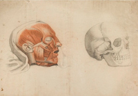
Why were the anatomical drawings so evidently important to Hosmer? If these drawings primarily reflect visual sources connected to medical rather than artistic education, what role could these images have played in her artistic career? As we will see, many of Hosmer’s early supporters pointed to her scientific training to silence doubts about a woman sculptor’s technical knowledge and bodily fortitude—a defense of her work which Hosmer may have underscored by displaying her anatomical studies. The unconventional training reflected in these drawings may also have been appealing to the artist who would become Hosmer’s mentor once she arrived in Rome: John Gibson (1790–1866), a Welsh neoclassical sculptor who had studied anatomy under a surgeon in Liverpool between 1813 and 1816.[41] Gibson’s working-class origins made him an unlikely candidate for academic training; like Hosmer, he had looked elsewhere for anatomical instruction. Gibson vividly recalled his first experience observing a dissection and making drawings while the surgeons worked, and he later joined local medical students “to rob dead bodies from their graves” in quest of anatomical specimens from the “public burying places.”[42] Some of Gibson’s surviving sketches from this period resemble not plates from medical textbooks but rather still-life drawings of dead bodies and body parts. One study in black and red chalk, for instance, depicts a head from which the skin has been removed, cradled in a crumpled white cloth (fig. 12). Its mouth gapes slightly open, revealing a row of small teeth, and the head tilts away from the viewer to rest upon an invisible surface. Numbers inscribed on various muscles, likely corresponding to a key on another sheet, subtly mark the artist’s scientific interest in the head. Gibson’s sketches suggest his observations of dissection more explicitly than do Hosmer’s, but both are distinct from La Farge’s studies of the nude. The experience of medical education which Gibson and Hosmer shared might well have contributed to the accomplished sculptor’s interest in his new pupil.
Though their visual sources lie in a medical publication, Hosmer’s drawings also make clear that her aspirations and sensibilities were not purely scientific. The three figures which Hosmer selected from the Anatomie élémentaire are distinct within that overall corpus of images. Bourgery and Jacob’s illustrations juxtapose multiple conventions of anatomical representation, ranging from the idealized écorché to graphic depictions of dissection and surgical procedure that testify to a more visceral engagement with living and dead bodies. Plate 12 of the Anatomie élémentaire, for instance, includes not only a central écorché figure in elegant contrapposto but also a more vivid illustration of a cutaway throat and shoulder, shown on the apparently living body of a man (fig. 13). Plate 18, demonstrating the organs of the abdomen, is explicit about the postmortem reality underlying the representation, as ragged flaps of tissue and muscle hang beneath each opened cavity in the center row of images (fig. 14). The lower-left vignette in plate 19 even includes the pins and clips inserted to tug apart various organs and render visible deeper layers of structure, bringing the process and apparatus of dissection into the image itself (fig. 15).
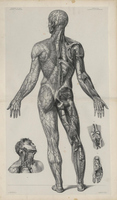
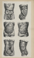
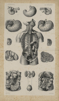
Bourgery and Jacob’s plates reflect a moment of transition in the discipline of anatomy and its visual culture. In historian Melissa Lo’s words, these pictures are “caught . . . between the binary system of art and medicine” and “symptomatic . . . of the epistemic rupture between the classical and the modern.”[43] This rupture, of course, extended far beyond a single publication, even one as capacious as Bourgery and Jacob’s project. As art historian Lyle Massey demonstrates, new kinds of anatomical images had gained epistemological primacy toward the end of the eighteenth century by “shun[ning] the aesthetic dimensions and classicizing ideals endemic to older anatomical traditions.”[44] The emerging norms of scientific and medical imagery dictated that “bodies [were] reduced to individual parts, and displayed as though in the process of being dissected,” stripped of the “tropes of lifelikeness” and other visual fictions.[45] The Anatomie élémentaire and the larger Traité complet were sprawling, unstable attempts at reconciliation of the classical, artistic ideal and the modern, scientific specimen as competing subjects of anatomical description and depiction in the mid-nineteenth century.
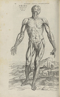
Hosmer’s anatomical plates, however, remain firmly rooted in the “system of art,” the “classical” image traditions of anatomy, and the ideal body as its subject. The stances and gestures of the Bourgery and Jacob figures which Hosmer chose to copy had first appeared almost three centuries earlier, in the work of the pioneering early modern anatomist Vesalius.[46] Illustrated with woodcuts that may have been created by multiple artists connected with the workshop of Titian (ca. 1488–1576), Vesalius’s De humani corporis fabrica libri septum (1543) (hereafter the Fabrica) includes several écorché figures from which Bourgery and Jacob’s, and thus Hosmer’s, images are derived.[47] One of the myological plates of the Fabrica, for instance, shows a flayed figure in contrapposto, with the index finger of its proper left hand pointing downward and its right held open, palm up (fig. 16). Similar figures appear in many subsequent anatomical publications from the sixteenth century onward, and Hosmer’s écorchés stand comfortably within this lineage.
Yet by the time Hosmer made her drawings, these Vesalian “muscle men” were no longer a universally accepted norm of anatomical representation.[48] By the end of the eighteenth century, physicians had begun to deride Vesalius and his followers for their “sturdy and active figures, with a ludicrous contrast of . . . active limbs, combined with ragged muscles, and naked bones, and dissected bowels,” in the words of the eminent Scottish surgeon John Bell, writing in 1794.[49] Anatomical illustrations, believed Bell and his allies, should be purged of these conceits and extracted from the fanciful scenery of trees, rocks, and classical ruins through which stroll Vesalius’s figures. Instead, anatomical specimens should appear in images as if under the surgeon’s knife, allowing clinical empiricism to replace the aestheticized naturalism of classical sculpture and pastoral landscape.[50] Rather than understanding art and anatomy as partners in the shared enterprise of producing and communicating knowledge about the human body, Bell described “a continual struggle between the anatomist and the painter; the one striving for elegance of form, the other insisting upon accuracy of representation.”[51] By 1850, many anatomical atlases and illustrations reflected this splintering of disciplinary conventions. It was not the idealized écorchés in Bourgery and Jacob’s treatise but instead their illustrations of bodily fragments and surgical procedures which reflected mid-nineteenth-century understandings of medical visualization.
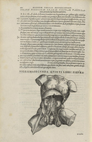
Why might Hosmer have chosen an antiquated mode of depiction when she had access to more contemporary models in the very same set of lithographs? Perhaps she did so because Vesalian anatomy was not simply a set of images but an epistemological proposition, reliant upon the idea that art and science were essentially allied modes of representing and understanding the human body. Hosmer—who wrote with equal enthusiasm to Cornelia Crow in 1851 about one of her first bas-reliefs in marble and her at-home dissection of a pet snake named Eve—likely found that belief powerfully appealing.[52] The visual influence of ancient sculpture on many of Vesalius’s represented bodies also offered Hosmer an opportunity to communicate her neoclassical tastes and sculptural ambitions. The Fabrica frequently depicted internal musculature and organs as if demonstrating dissections of famous works of classical art (fig. 17), such as the so-called Belvedere Torso, a marble fragment dating to the first century BCE. (Bell singled out for special mockery the fact that Vesalian anatomists “borrow the celebrated Torso [Belvedere] for putting their bowels into.”[53]) By a strangely circular logic, Vesalius also encouraged readers of the Fabrica to perform public dissections upon actual bodies that corresponded to ancient Greek canons of proportion and could thus serve as normative standards of comparison.[54] Visual fictions and careful selection of anatomical subjects helped to construct the idealized, universalizing body that was a central figure of early modern humanism. Hosmer’s carefully copied Vesalian figures positioned the young sculptor as an inheritor of this conjoined tradition of anatomical art and artistic anatomy.
Whose Bodies?
Unlike the medical images gaining epistemological currency in the mid-nineteenth century, Hosmer’s Vesalian bodies disguise the messy, visceral fact of dissection, cloaking the mangled flesh of a once-living person in aesthetic convention. As art historian Glenn Harcourt observes, the “idealized, classical forms of [Vesalian] figures, the fact that they do not read as actual cadavers” elides the unsettling reality that “anatomical knowledge is constituted through the violation and destruction of its proper object in practice,” the human body.[55] Since the early modern period, these visual fictions had been part of a “representational strategy that at once belies and elevates the socially problematic practice of physical violation characteristic of the real production of anatomical knowledge.”[56] Vesalian anatomy served not only an artistic or a scientific end but also a social purpose.
That social purpose remained critical in the time and place in which Hosmer produced her drawings. Despite the growing importance of dissection in medical education in the nineteenth-century United States, the practice remained socially unsanctioned, even morally taboo. The only cadavers that were legally available for dissection in most states were those of executed criminals, who could be subjected to the additional indignity of postmortem punishment. In Missouri, a state law enacted in 1835 made one other category of persons available for anatomical study: “the body of a slave, with the consent of his owner,”[57] which historian Daina Ramey Berry terms “postmortem commodification.”[58] Given the scarcity of legally dissectible cadavers, many nineteenth-century doctors in the United States also resorted to illicit means to procure anatomical subjects. Postmortem theft posed the greatest danger to racialized, impoverished, and socially marginal subjects: vulnerable people without kin to claim them and communities without power to protest the violation of their resting places.[59] Grave robbers in the pay of medical schools (and sometimes, doctors and medical students themselves) targeted cemeteries in Black neighborhoods as well as so-called potter’s fields and other burial grounds that primarily received the bodies of the poor.[60] As art historian Naomi Slipp writes, this trade in cadavers meant that “the furthering of medical science”—and artistic production—“relied on unequal power relations that allowed for the exploitation of the country’s most disenfranchised citizens.”[61] The unsettling politics of dissection are frequently “buried beneath ideal exteriors,” like those of Hosmer’s figures.[62]
To understand the social meaning of Hosmer’s anatomical drawings, we must pose the questions that Vesalian anatomical conventions seek to repress. As art historian Keren Rosa Hammerschlag argues, despite the importance of race and class in determining whose bodies were used for anatomical study, nineteenth-century anatomical atlases and illustrations “are rarely viewed through the lens of race” or other categories of social positionality.[63] As we recall, many anatomical figures are depicted without skin, rendering racialized identity illegible; the convention of posing anatomical subjects as “Greco-Roman statues” suggests their presumptive whiteness.[64] But underneath the layers of representational tradition and classicizing aesthetics, whose bodies were these? If the visual strategies of Vesalian anatomy serve to disguise the mechanics and implications of its real practice, what aspects of Hosmer’s real experiences in St. Louis are recoverable?
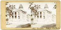
Such questions are particularly significant in Hosmer’s case, because the artist’s decision to attend the medical college superintended by McDowell placed her at the center of vociferous, sometimes violent, contestations over anatomy’s permissibility and meaning in a society defined by racial inequality. By the 1850s, McDowell possessed a grim reputation for his and his pupils’ practice of illegally exhuming corpses from St. Louis’s graveyards to procure subjects for dissection. On at least two occasions, mobs ransacked the medical college, searching for the missing corpses of kin, friends, and neighbors.[65] McDowell reportedly stockpiled arms and ammunition in the imposing college building at Eighth and Gratiot Streets, in constant readiness for attack by enraged citizens (fig. 18).[66] Particularly afflicted by the anatomist’s rapacity for dissection subjects were St. Louis’s enslaved and people of color as well as the city’s large population of German immigrants. As McDowell’s niece later recalled, Black and German St. Louisans so despised the doctor that they would “cross the street” to avoid walking past the medical college where Hosmer spent her days with McDowell.[67]
McDowell was hardly alone among nineteenth-century doctors in resorting to illegal methods to procure anatomical specimens, nor in his use of the bodies of racialized subjects. Yet the St. Louis anatomist’s evident unconcern for the dignity of the dead among the city’s Black and immigrant population may also reflect his own deeply held beliefs about racial hierarchy. In December 1859, as the United States lurched toward civil conflict over slavery, McDowell published an invective against the abolitionist minister Henry Ward Beecher. His letter, reprinted in newspapers across the South and Midwest, used his anatomical experience to support the view that inequality between white and Black people was just, natural, and immutable: “There is no identity of origin, no equality in mental and physical organization. . . . The negro has been what he is for four thousand years, and will be the same for four thousand years to come.”[68] McDowell’s prejudices against immigrants and Catholics were almost as vitriolic as his anti-Black racism, for he was an enthusiastic member of the Know Nothing Party and, as one sympathetic newspaper described him in 1856, “a gallant champion of the great principle that Americans should rule America.”[69] Paradoxically, however, the doctor and his pupils likely gained their knowledge of the human form from the dissection and observation of precisely those bodies which McDowell deemed to represent inferior humanity.[70] The doctor closed his letter to Beecher by proclaiming his own willingness to use violence in defense of human bondage: “I have wielded the knife in surgery for profession . . . [but] when my country is destroyed by heartless fanatics, excited to the deeds of bloodshed, the knife . . . will be whet in justified vengeance.”[71] With the outbreak of the American Civil War, McDowell joined the proslavery cause as the Confederate army’s Surgeon General of the West.[72]
Given the source of many cadavers among the enslaved, the poor, and within Black and immigrant communities, debates over dissection became touchstones for broader conflicts over race, self-possession, and the value of human life in the antebellum United States. The large-scale traffic in stolen bodies meant that the threat of dissection also represented the threat of commodification: the reduction of a human being to an object for sale. The Daily Messenger of Hannibal, Missouri, for instance, reported in 1853 that “a number of young men,” happening upon “a seedy-looking individual perfectly insensible from the effects of liquor,” decided to “bag the drunken loafer and sell him to the doctors at a medical college” for five dollars, as a specimen for dissection.[73] Or, as an article in the Waverly, Missouri, Saturday Weekly Visitor noted in 1860, an “old man . . . who is owing a physician a bill of thirty dollars, and unable to pay it, has deeded to the doctor his body for dissection after his demise.”[74] The fact that none of the actors in these small dramas are explicitly racialized in the newspaper reports strongly suggests that all were white. It is impossible to know how much truth these stories contain, but the narratives raised the possibility that dissection might threaten to consign white subjects to a condition of dispossession like that of the enslaved, who could be legally bought and sold before and after death.
Antislavery advocates, for their part, used the widespread distaste for dissection to argue that enslavement was an equally repugnant form of bodily theft. As one writer from Auburn, New York, fumed in William Lloyd Garrison’s Liberator in 1846, the reported theft of “the body of a white man . . . by some young doctors for dissection” had produced “the most intense and thrilling excitement” and a massive search for the missing body, but “at about the same time, a fugitive slave from Virginia” was recaptured by slave hunters.[75] The writer continued: “In the middle of the day, [the fugitive] was borne out of the country . . . seen by hundreds of those who had been ready to ‘lynch’ the grave robbers.”[76] The spectacle of a community up in arms on behalf of a dead white body, and indifferent to the fate of a living Black man, offered grim proof of the disparate value of life in the nineteenth-century United States.

Did Hosmer witness dissections of the bodies of Black St. Louisans, immigrants, or other racialized subjects during her studies in the city? Did she gain the expertise displayed in her anatomical drawings from such observations? To my knowledge, no evidence survives that would offer conclusive answers to these questions, but it seems probable that she did. She could hardly have remained unaware of McDowell’s avowedly racist views or his belief in the anatomical foundations of human difference. Her residence in Missouri during the final, turbulent decade of sectional conflict prior to the Civil War would also have exposed her to other aspects of the politics and lived reality of racial hierarchy and racial capitalism. St. Louis was both a Western and a Southern city, whose slave markets profited from the trade in enslaved people along the Mississippi to the Deep South.[77] Residing with Wayman Crow and his family would also have brought Hosmer into intimate, daily contact with enslaved people—probably for the first time, since she had lived until then in Massachusetts, where slavery had been judicially abolished in the 1780s.[78] Crow enslaved one man and two women in his household in 1850, the year in which Hosmer lived there (fig. 19). Hosmer’s later letters to the Crow family, in which she occasionally sends greetings to the three enslaved domestic workers, reveal that their names were Tom, Jane, and Emily. According to the 1850 slave schedule, which records the gender and age but not the names of enslaved people, either Jane or Emily was then nineteen years old, a year younger than Hosmer herself.[79] When Crow manumitted Tom, Jane, and Emily in 1853, Hosmer expressed her approbation, writing to him of “that gift of freedom, which was truly a grand thing. If all the masters would do the same what a glorious change there would be.”[80] Yet even as Hosmer praised Crow and expressed her desire for universal manumission, she did so in terms that reflected her view of enslavement as essentially paternalistic and benevolent. She wrote that she expected to see all the newly freed people still working in Crow’s household whenever she visited, since “they wouldn’t leave you if they could . . . for they must consider themselves bound by the strongest chains of love & gratitude.”[81] To Crow’s daughter Cornelia, Hosmer wrote: “So Jane & Tom & Emily . . . are going on their own books. It was a grand thing for your father to give them their freedom but I dare swear they are not one half so free or so happy as of yore.”[82] Hosmer echoes a trope that would be taken up by many white supremacists after the Civil War and emancipation: that Black Americans were better off as slaves than free.
Hosmer’s residence in St. Louis in 1850 and 1851 laid a foundation for her lifelong connection with the Crow family, and particularly her relationship with Wayman Crow, who would become her most generous and determined advocate in the United States. The economics of sculptural production meant that Hosmer needed a deep-pocketed patron like Crow, since carving in marble necessitated capital to purchase the costly material, sometimes long before she could expect to sell the finished work. She wrote to Crow from Rome in October 1854, confessing that she had “two or three bills coming due for marble” and appealing to his entrepreneurial spirit: “You, dear Mr. Crow, know that one must have a good capital to begin business at all, much more to begin with advantage. It is exactly the same thing in Sculpture. A painter has no expense of the kind, he buys his canvasses and paints which cost little or nothing & he is made, but from the time a sculptor begins he finds that without funds he is at a standstill.”[83] Perhaps unconscious of the irony of addressing such a sentence to a former slaveholder, she wrote to her patron about a possible commission in 1859: “All that my head or hand can do is yours at command as I consider that you pretty well own me.”[84] Hosmer’s financial dependence on Crow’s support meant that her professional success was in part enabled by the stolen labor of human beings on Crow’s properties in Missouri and Kentucky, on which he had enslaved at least eight people.[85] Her entanglement in such transactions is worth noting not because it makes her unique but because it does not. Instead, her indebtedness to Crow reflects what art historian Caitlin Meehye Beach describes as the systemic dependence of “the sculptural enterprise and medium” on “modes of commerce and commodification” in the nineteenth century.[86] Like many of her contemporaries, Hosmer forged her career within “an Atlantic world of artistic production, patronage, and commerce” contoured by racial capitalism.[87]
We may be tempted to dismiss the proslavery views expressed in Hosmer’s letters to Crow and his daughter as flattery of the artist’s most important patron and her closest friend, yet archival evidence suggests that Hosmer expressed similar opinions even in circles in which such views made her an outlier. Many of Hosmer’s acquaintances and supporters were prominent figures in the antislavery cause: the editor and writer Lydia Maria Child, the British actress Fanny Kemble, and the upper-class Bostonian Sarah Shaw, whose son Robert Gould Shaw would die alongside Black troops at the Battle of Fort Wagner in 1863. Hosmer’s own father is noted on a list of donors to the National Anti-Slavery Standard, the weekly newspaper of the American Anti-Slavery Society, in 1846.[88] Despite her personal affection for Hosmer, Child chided the sculptor for her violent expressions of anti-abolitionism. Writing to Hosmer in September 1860, Child described a visit to the poet John Greenleaf Whittier, an “old Anti Slavery friend” and “one of those whom you wanted to have hung, alongside of me.”[89] Nor was Child in any doubt as to the geographic origin of Hosmer’s thinking: “If you had had your will, ‘Missouri Ruffian’ that you are! and had exterminated the abolitionists, let me tell you, you would have destroyed the wheat of the country, and left nothing but the chaff.”[90] Half mockingly, she offered to send Hosmer “a bowie knife, bearing the motto ‘Death to abolitionists,’” so that she could settle disputes with her rival artists “Missouri fashion,” likely a reference to violence by proslavery Missourians who had crossed the border into Kansas after the passage of the Kansas-Nebraska Act of 1854.[91] Child’s letter, written almost a decade after Hosmer left St. Louis, attests to the enduring impact of her experiences there.
Returning to Hosmer’s anatomical drawings, the sculptor’s education and experiences in St. Louis suggest a necessary reframing of the significance of Vesalian anatomy for artists and audiences in the mid-nineteenth-century United States. Like many anatomical images in the Vesalian tradition, Hosmer’s drawings disavow their practical origins in dissection, eliding the physical violence and moral violation necessary to produce them. In doing so, these drawings also disavow their “social origins” in the bodies of racialized and marginalized people.[92] If, as art historian Charmaine Nelson argues, “the always already white marble of neoclassicism persistently refused the representation of the black body at the level of skin/color,” the anatomical study that preceded sculpture both drew upon and disavowed nonwhite bodies at a level below the skin.[93]
Aesthetic Medicine
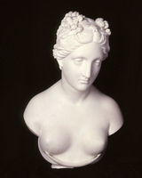
Significant as Hosmer’s anatomical drawings were to her, their purpose was ultimately to prepare her for something else: a career making figurative sculpture in marble. Anatomical study was critical to Hosmer’s artistic training, but did it influence her sculptures and their reception? After Hosmer returned to Watertown from St. Louis, she completed her first neoclassical bust in marble, Hesper, the Evening Star (1852), a female personification of the heavenly body with sleep-inducing poppies wound in her hair (fig. 20). Writing wittily in The New-York Tribune of “A New Star in the Arts,” Child praised the bust’s convincing corporeality, describing “the swell of cheek and breast . . . like pure, healthy young flesh” and the “muscles of the beautiful mouth [which] are so delicately cut it seems like a thing that breathes.”[94] Though Child and Hosmer might have argued over the opinions on slavery that Hosmer acquired in St. Louis, they agreed on the value of the anatomical knowledge she gained there. Child attributed the success of the sculpture to Hosmer’s serious study of “flesh” and “muscles,” writing that “Miss Hosmer places a just value upon scientific rules. Few artists among us have such an accurate knowledge of anatomy.”[95] A few months later, a similar account of one of Hosmer’s early sculptures appeared in the “Editor’s Table” of Godey’s Lady’s Book, at the time the most widely circulated magazine in the United States. Likely written by the publication’s editor, Sarah Josepha Hale, the piece cited McDowell’s praise for Hosmer’s “rare proficiency” and his opinion that “the now world-renowned [Hiram] Powers” had not “made such progress in anatomy as Miss Hosmer had attained at the time of her departure” from his medical college.[96] The Godey’s article offered further proof of Hosmer’s skill in representing the human form by describing her marble portrait medallion of McDowell, which was “as large as the natural head,” with the features “admirably preserved.”[97] Hosmer, the writer concluded, “has shown the superiority of the female mind in the study of anatomy, thus pointing to woman’s true profession in the sciences, viz., the medical.”[98]
The favorable reception of Hosmer’s earliest sculptures rested upon the anatomical achievements and scientific expertise of the young sculptor, perhaps even more so than upon her taste, style, or subjects. Both Child and Hale were among the antebellum period’s determined advocates of expanding educational opportunities for women, which may explain their close attention to the fruits of Hosmer’s anatomical training. Their praise of Hosmer in medicalized terms—as a sculptor of “healthy” flesh, a scientist in stone—also suggests what kind of education they considered important, reflecting the priorities of a nascent movement to incorporate physical education, health sciences, and even anatomical study into women’s schooling. This educational-reform movement was in fact partly responsible for the US circulation of the same anatomical images which Hosmer had reproduced during her studies in St. Louis. As we have seen, she likely copied her three figures from plates originating in Bourgery and Jacob’s Anatomie élémentaire. Among the most enthusiastic promulgators of Bourgery and Jacob’s images in the United States was the physician, lecturer, and educator Lambert, who published several mass-market textbooks and folios containing these anatomical images in the early 1850s (see figs. 8, 9). Lambert was not affiliated with any medical school but instead served as the “Professor of Anatomy and Physiology in Pittsfield (Young Ladies’) Institute,” a women’s school in the Berkshires that catered to daughters of the New England elite.[99]
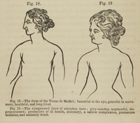
Lambert’s “physiology” texts were not intended to train future doctors but instead aimed at laypeople, particularly women. His works formed part of a developing corpus of literature supporting the period’s “physical culture and hygiene movements, which sought to popularize good health.”[100] In the opinion of Lambert and other proponents of “physical culture,” the human body bore the legible signs of “good health” or its absence, making anatomy not only a scientifically descriptive but also a socially prescriptive endeavor. Lambert’s treatise Popular Anatomy and Physiology opens with two plates showing Bourgery and Jacob’s myological figures. The book’s subsequent discussion of bone, muscle, and organ structure is centrally concerned with the use of anatomical representation to influence social norms and individual behavior. A pair of images demonstrates the anatomical effect of “female dress, as usually worn,” by comparing the “form of the Venus de Medici; beautiful to the eye, graceful in movement, healthful, and long-lived,” with the wasp-waisted torso produced by corsetry, which Lambert describes as “ungraceful, disproportioned; productive of ill health, deformity, a sallow complexion, premature wrinkles, and untimely death” (fig. 21).[101] (The visual resemblance between the illustration and the famous Venus de’ Medici, a first-century BCE marble sculpture, is somewhat approximate.) Lambert’s medical text supported an emerging movement for “dress reform” in the nineteenth-century United States, whose adherents decried the destructive effects of lacing and whalebone on women’s bodies.[102] Though many dress reformers were early feminists, the movement also included fellow travelers like Lambert, who were less interested in revolutionizing women’s traditional roles than in ensuring that women were healthy enough to fulfill them.
These images of Venus and her corseted counterpart operate at the same unstable boundary between the ideal and the real, the scientific and the sculptural, where we might locate Vesalius’s Belvedere Torso spilling its guts, or Hosmer’s carefully posed écorchés. Nineteenth-century dress reform and Vesalian anatomy each assumed commensurability between actual human bodies, anatomical images of human bodies, and ancient sculptures. As art historian Hallie Franks writes, the use of classical art in dress-reform texts “positioned sculpture[s] as aspirational models for real bodies,” much as the earlier Vesalian tradition had positioned antique sculptures as normative human forms.[103] Indeed, even the most trenchant critics of Vesalian anatomy and its classicizing aesthetics tended to accept that ancient sculptures reflected some degree of anatomical truth. Though a Vesalian skeptic, the eighteenth-century surgeon Bell accepted that figurative sculpture made by the Greeks, who saw “in their women, the purest models of female beauty” and “in their young men, the grandest displays of the manly form,” were anatomically correct.[104] The ideal proportions and developed musculature of Greek sculptures were accurate representations of those ancient subjects, Bell conceded, but they were hardly adequate models for the bodies of “the moderns [who] have come poorly after” or the modern artists who “cop[ied] coldly those half-animated forms” of ancient art.[105] The problem with using antique statues as anatomical subjects lay in the present rather than the past. Ancient Greek sculptures, and the ancient Greek bodies which those sculptures represented, reflected an acme of human embodiment long vanished.
The concept of degeneration from a former ideal of physical perfection incarnated in classical Greece had deeply racialized implications—as did the emerging discourses around women’s physical health and hygiene.[106] Just as he had copied the Bourgery and Jacob plates from French publications, Lambert did not originate the comparative images of the Venus de’ Medici and the corseted body which appeared in his physiological texts. As Franks has shown, Lambert reproduced images that had begun circulating in the United States during the 1840s, in publications by the phrenologist Orson Squire Fowler (1809–87).[107] Like other exponents of this popular pseudoscience, Fowler held that innate links existed between physical form, human intelligence, and moral character. While phrenology is usually understood as the belief that such traits were legible in the skull, Fowler believed that many aspects of human physiology possessed this potential for signs of superiority or inferiority. In his view, artificially narrowed waists augured disease, weakness, deformity, and even sexual immorality.[108] As with most phrenological discourse, Fowler’s warnings about the evils of corsetry were linked to ideas about racial hierarchy. He predicted that lacing “will kill all fashionable women and their children, and leave our square-formed, broad-shouldered, and full-breasted Irish and German women alone for wives and mothers.”[109] Fowler played on fears of racial degeneration produced by growing numbers of immigrants to the United States, many of them fleeing famine in Ireland and the unrest produced by the revolutions of 1848 across Europe. Speaking, presumably, to an elite white audience, he fumed that women’s love of dress had “already alarmingly deteriorated our race in both physical and intellectual stature, and unless checked, will soon destroy it.”[110] His physiological text constitutes an early contribution to eugenic discourses and fears of “race suicide” that would gain ground over the subsequent decades of the nineteenth century. Similar plates comparing the waistline of the Venus de’ Medici and the lady of fashion were recirculated in eugenics texts as late as 1904.[111]
Hosmer was almost certainly aware that anatomy and classical sculpture were entangled in these period discourses concerning women’s health. The sculptor’s own upbringing was an early example of physiology playing an essential role in a woman’s education. Harriet’s two younger brothers and her mother had died of tuberculosis between 1832 and 1836; her elder sister, Helen, succumbed to the same disease in 1842.[112] Hiram Hosmer, himself a physician, placed his surviving daughter on a “physical fitness regimen that included hiking and hunting on the land around their home and swimming in and rowing on the Charles River.”[113] Given that corsets were believed by their critics to constrict the flow of air through the lungs and aggravate or even cause tuberculosis, it seems unlikely that Hosmer’s father wished her to wear one. Throughout her career in Rome, Hosmer continued to ride, hunt, and walk, sometimes provoking disapproval for her willingness to venture out alone for exercise.[114] But her exemplary physical health was also essential to her professional success, for her robustness helped convince the public that a woman could undertake the strenuous labor of chiseling in marble. As Culkin writes, early criticism of Hosmer’s works often described the artist’s “unusual, athletic upbringing,” which had granted her “the physical and emotional wherewithal to become a sculptor.”[115]
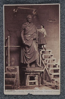
Another point of contact between Hosmer and these medical and educational discourses was the question of women’s dress, an issue which engaged the artist throughout her life. In the first decades of her career, she wore what she referred to as a “Zouave costume” of loose-fitting trousers and short jacket while working in her studio and even occasionally donned men’s clothing in public.[116] Between 1860 and 1862, for instance, she posed for a photograph in an outfit composed of beret, blouse and jacket, belted, knee-length skirt, and what appear to be breeches and stockings, while at work upon a monumental statue of Thomas Hart Benton (1782–1858), the powerful US senator who had represented antebellum Missouri (fig. 22). She wrote to Wayman Crow that she had dispensed with long skirts because she was “not intending to break [her] neck upon the scaffolding [of the Benton statue] by remaining in petticoats.”[117] However, Hosmer did not endorse dress reform’s potential feminist implications: As she wrote to Crow in 1859, she disdained “bloomerism & that view of woman’s rights.”[118]
Later in her career, however, Hosmer became a vocal advocate of dress reform from an ideological as well as a practical standpoint. She appeared publicly in St. Louis in 1888 in a corset-less garment of “black silk, the folds of which were so ample as to be suggestive of the ancient Roman toga,” which was “but slightly confined at the waist by a broad girdle.”[119] A reporter for the St. Louis Globe-Democrat described Hosmer’s dressed body in terms that might have been lifted from one of Lambert’s or Fowler’s texts celebrating the “natural” waistline: “The contour of form produced was like that of a Roman matron in Grecian draperies.”[120] In her own remarks to the reporter, Hosmer cemented this connection by extolling “Grecian models” as the “acme of excellence” and observing that though “the Venus de Medici is as large about the waist as about the chest,” most fashionable women would “weep” to appear in the “unconfining, flowing draperies” of classical sculpture.[121] She further justified her disapproval of corsetry by recalling one particularly vivid experience from decades prior, in McDowell’s anatomical theater: “Dr. McDowell once lectured on the form of a lady who had died from tight lacing. The ribs, instead of diverging diagonally downwards, had been cramped up until they interlaced. This had forced the upper organs up and the lower ones down, until death had resulted.”[122] With striking precision, Hosmer echoed the period’s phrenological, physiological, and eugenic texts, elevating the Venus de’ Medici as a model of feminine beauty, condemning fashionable dress as a danger to women’s health, and marshaling anatomical evidence to support her claims.
Hosmer’s particular brand of feminism, with its classical ideal of the normative woman and its affinities with early eugenics, belied the movement’s antebellum origins in close alliance with abolitionism. Perhaps unsurprisingly, when the US feminist movement split over the issue of Black suffrage, Hosmer supported the National Woman Suffrage Association, led by Susan B. Anthony (1820–1906) and Elizabeth Cady Stanton (1815–1902), which opposed the Fifteenth Amendment to the US Constitution granting Black men the right to vote. During a visit to New York City in the late 1860s, Hosmer called upon Anthony and subscribed to The Revolution, the weekly published by Anthony and Stanton.[123] Hosmer’s subscription would have exposed her to the newspaper’s sometimes explicitly anti-Black and anti-immigrant arguments for (white) women’s suffrage. As Stanton exhorted her readers in the issue of April 29, 1869: “American women of wealth, education, virtue, and refinement, if you do not wish the lower orders of Chinese, Africans, Germans and Irish, with their low ideas of womanhood[,] to make laws for you and your daughters . . . demand that woman, too, shall be represented in the government!”[124] Stanton’s and Hosmer’s feminism was, emphatically, white feminism.
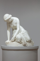
Hosmer’s early work in marble suggests that these physical and social ideals shaped the construction of her neoclassical female bodies. As Hosmer wrote to Cornelia Crow in December 1852, a month after her arrival in Rome, she had begun her studies with Gibson by copying from the antique. She announced proudly: “I commenced my campaign with the Venus of Milo, in fact have not yet finished it & am getting along very nicely with it.”[125] Like the Venus de’ Medici, the Venus de Milo, dating to the second century BCE, is a monumental Hellenistic marble sculpture of a female figure, displaying a plump, muscular torso and ample waist.[126] Hosmer’s first full-length, life-size marble sculpture, Oenone (1854–55) (fig. 23), explicitly draws upon the classical model which Gibson had set her to emulate. Representing the wood nymph whom Paris abandoned for Helen, the seated figure droops mournfully over the Trojan prince’s discarded shepherd’s crook. With Oenone’s drapery slipping around her waist to reveal her bare torso, the sculpture pays homage to the Venus prototypes. Oenone’s gracefully sloping shoulders, wide chest and waist, and soft, smooth flesh reflect the qualities which neoclassical sculptors and dress reformers alike prized as the “acme of excellence” in female form.
In subtler ways, too, Oenone represents a meditation on the measure of women’s physical beauty. Hosmer, an avowed admirer of Alfred, Lord Tennyson (1809–92), likely took inspiration from the poet’s Oenone, published in 1833.[127] Tennyson’s verses are an angry lament in the voice of the scorned nymph, who recounts her beloved’s choice of Aphrodite in the contest of beauty between the goddess of love and her rivals.[128] The victorious Aphrodite bribed Paris with the “fairest” wife in Greece, but, as Oenone apostrophizes: “Am I not fair?”[129] Abandoned by her lover, Oenone contemplates revenge “as a mother / Conjectures the features of her child / Ere it is born.”[130] Yet for the nymph, there will be no true motherhood: “Never child be born of me, / Unblest, to vex me with his father’s eyes!”[131] Though art historian Joy S. Kasson suggests that the textual underpinnings of Hosmer’s subject recast the abandoned woman as a figure of potential “strength” and “retribution,” it is also possible to understand Oenone as a tragic figure in more conventionally gendered terms.[132] Denied a woman’s destiny in love, marriage, and motherhood, Oenone descends into madness and hatred: a failure of reproduction that ends in self-destruction. In this sense, the thematics of Hosmer’s Oenone resonate with the forebodings of physiologists and eugenicists, with fears of falling birthrates and race suicide. Denied their reproductive capacities by the desertion of faithless men or their own mistaken adherence to the dictates of fashion, beautiful women without children were both villains and victims of tragedy.
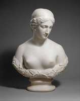
We may read Hosmer’s choice to sculpt an allegory of reproductive failure as either strategic or ironic, given the sculptor’s own rejection of heterosexual marriage and childbearing. As she wrote to Wayman Crow in 1855, noting several weddings among their mutual acquaintance: “You see everybody is being married but myself. . . . An artist has no business to be married—for a man it is all well enough but for a woman on whom matrimonial duties & cares weigh more heavily it is a moral wrong, I think, for she must either neglect her profession or her family, becoming neither a good wife & mother [n]or a good artist.”[133] Hosmer’s reasons for refusing to become a “wife & mother” were both professional, as she stated to Crow, and personal, since her life included intimate, romantic, and avowedly conjugal relationships with other women.[134] Hosmer often addressed her difference from conventional motherhood by evoking the discourse of maternity in relation to her sculptures, playfully but consistently casting them in terms of biological reproduction. She referred to a marble bust of Daphne (fig. 24) in a letter from 1854 to Wayman Crow as her patron’s “marble daughter,”[135] and to Cornelia described the bust as her own “first child.”[136] Writing in June 1853 about Cornelia’s impending marriage, Hosmer mused that her friend should give birth to two daughters: “The first daughter will be Cornelia & the second daughter will be Hat for short.”[137] For herself, though, Hosmer confessed: “I like marble babies best. . . . You will see one of my babies before I see one of yours.”[138] The sculptor’s language both accommodates and reframes the notion that women were naturally suited to maternal roles. Yet such “discursive play with sculptural ontology” also racialized Hosmer’s sculptures by placing them in biological kinship with the white artist and her white patrons.[139]
Of course, the waistline was not the only locus at which neoclassical sculpture overlapped with the discourses of racial embodiment.[140] Neoclassical sculpture reflected an enduring link between the whiteness of stone and the whiteness of skin, between chromatic and biological whiteness. Hosmer certainly understood these associations. She wrote to Cornelia Crow around 1853, explaining her delay in dispatching Daphne from Rome as a gift to the Crow family: “Her little face was not quite clear, wanted a draught of sarsaparilla to purify it, & so . . . I ordered another one to be cut.”[141] Hosmer’s reference to sarsaparilla, a nineteenth-century patent medicine believed to cure skin complaints, suggests that veins or spots had appeared in the white marble during carving. The medicalized language is surely tongue-in-cheek, but the metaphor illustrates Hosmer’s assumption that her sculptures’ pale surfaces should mimetically represent unblemished white skin. The same sense of intimate connection between real and sculptural bodies, between medical science and art, that produced Hosmer’s horror at a human torso corseted out of resemblance to “Grecian ideals” led her to prescribe a pharmaceutical treatment for darkened stone and describe her parturition of “marble babies.” Hosmer, in her own way, was reproducing whiteness.
Anatomical study and neoclassical sculpture were bound together in Hosmer’s artistic formation, providing essential context for understanding both her career and the nexus of race, gender, and sculpture in her work and in the nineteenth-century United States. Reconsidering Hosmer’s anatomical training suggests a more complex and ambivalent story than was told by the artist herself or repeated by her critics and biographers. Hosmer surmounted the gendered barriers to anatomical study that hindered many nineteenth-century women artists: a key step toward her future profession in neoclassical sculpture. Yet we must look critically at the institutions, discourses, and networks which allowed her to do so, and the ideologies and loyalties which shaped her as a result. Keeping in view the anatomy of neoclassical sculpture—that which lies beneath its smooth surfaces—allows for a more thorough reckoning both with Hosmer’s practice and with nineteenth-century sculpture itself.
Acknowledgments
My sincere thanks to the editors of Nineteenth-Century Art Worldwide as well as the two anonymous reviewers for their thoughtful and generous engagement with this essay. I am indebted to Madeleine Murphy, access services coordinator at the Schlesinger Library on the History of Women in America, Radcliffe Institute for Advanced Study, Harvard University, for making Hosmer’s anatomical drawings available to me for study. I would also like to thank the Association of Historians of Nineteenth-Century Art for the opportunity to present an early version of this work at the 22nd Annual Graduate Student Symposium in Nineteenth-Century Art in March 2025. Jennifer Raab and Rachael Z. DeLue offered encouragement and generative questions during the development of the project. Finally, my gratitude to Siobhan Angus, Caterina Franciosi, Natalie E. Wright, and Joseph Mizhakiiyaasige Zordan for their invaluable feedback on this article.
Notes
[1] “The Studio of a Lady of Genius,” Vermont Journal (Windsor, VT), November 26, 1852, 1.
[2] The Vermont Journal also reported that Hosmer’s studio contained a “dried human heart,” a collection of insect specimens, and “numerous stuffed birds, pickled vipers, and skeletons of various small animals.” Where Hosmer obtained human remains, or whose remains they were, is not known, nor is the fate of the remains after her departure for Rome. “The Studio of a Lady of Genius,” 1.
[3] Kate Culkin, Harriet Hosmer: A Cultural Biography (University of Massachusetts Press, 2010), 28.
[4] One of the three anatomical drawings (corresponding to fig. 2 of the present article) is reproduced in Culkin, Harriet Hosmer. Culkin writes that “Harriet produced detailed, eerily accurate drawings of human muscle patterns, demonstrating her dedication to her work and talent as a draftsman.” Culkin, Harriet Hosmer, 17. Another drawing (corresponding to fig. 3) is reproduced in a short online feature by Melissa L. Gustin, curator of British Art at the National Museums Liverpool. See Melissa L. Gustin, “Harriet Hosmer: ‘The Queerest, Best-Natured Little Chap Possible,’” Stories, National Museums Liverpool, accessed January 19, 2026, https://www.liverpoolmuseums.org.uk/. Gustin does not describe the drawing itself but states: “After leaving school in 1849 Hosmer decided to pursue sculpture as a career, and went to medical school to study anatomy in St[.] Louis so she could more accurately depict the human figure.” Regarding the sculptor’s interests in science, art historian Buket Altinoba has recently discussed some of Hosmer’s later technological inventions, suggesting how important these interests remained throughout her career. Buket Altinoba, “‘Anybody Can Do Sculpture, It Is Purely Mechanical’: Harriet G. Hosmer and the Relation Between Sculpture, Reproduction and Machine Invention,” Sculpture Journal 33, no. 1 (March 2024): 29–46. Gustin has also written about Hosmer’s experimental approach to sculptural materials and engagement with nineteenth-century archaeology: Melissa L. Gustin, “‘Death, Like a Sculptor’: Archaeology, Materials and Text in Harriet Hosmer’s Pompeian Sentinel,” Sculpture Journal 33, no. 1 (March 2024): 47–70.
[5] McDowell privatized the medical school in 1857, detaching it from the state university and rechristening it Missouri Medical College. See Stephen Logsdon, “Joseph Nash McDowell, the St. Louis legend,” Archives and Rare Books (blog), February 10, 2022, Bernard Becker Medical Library, Washington University in St. Louis School of Medicine, https://becker.wustl.edu/; and James Walter Wilson, “Joseph Nash McDowell, M.D.,” Register of the Kentucky Historical Society 68, no. 4 (October 1970): 341–69. In 1899, Missouri Medical College was incorporated into the medical school of Washington University in St. Louis. Logsdon, “Joseph Nash McDowell”; and Wilson, “Joseph Nash McDowell,” 367.
[6] For a broader discussion of the role of anatomy in nineteenth-century art education in the United States, see Alan C. Braddock, “‘Jeff College Boys’: Thomas Eakins, Dr. Forbes, and Anatomical Fraternity in Postbellum Philadelphia,” American Quarterly 57, no. 2 (June 2005): 355–83; Naomi H. Slipp, “‘The Secret Figure’: Artistic Anatomy and the Medical Body in Nineteenth-Century American Culture” (PhD diss., Boston University, 2015); Naomi H. Slipp, “Thomas Eakins and the Human Écorché: Understanding the Body in Three Dimensions,” Sculpture Journal 24, no. 3 (2015): 333–80; and the essays in Teaching the Body: Artistic Anatomy in the American Academy, from Copley, Rimmer, and Eakins to Contemporary Artists, ed. Naomi Slipp, exh. cat. (Boston University Art Gallery, 2013).
[7] T. C. Lee, “Anatomy and Academies of Art II—a Tale of Two Cities,” Journal of Anatomy 236, no. 4 (April 2020): 582.
[8] Meredith Gamer, “The Smugglerius, Re-Viewed,” Sculpture Journal 28, no. 3 (November 2019): 334.
[9] Slipp, “‘The Secret Figure,’” 1.
[10] “Harriet Hosmer,” St. Louis Globe-Democrat, December 13, 1888, 8.
[11] Culkin, Harriet Hosmer, 16.
[12] Culkin, Harriet Hosmer, 16. See also Janice P. Nimura, The Doctors Blackwell: How Two Pioneering Sisters Brought Medicine to Women—and Women to Medicine (W. W. Norton, 2021); Stacy Weiner, “A Brief Timeline of Women in Medicine,” AAMC News, March 5, 2024, American Association of Medical Colleges, https://www.aamc.org/; and “The Path to Medical Coeducation in the United States,” Legacy Exhibit, Women in Health Sciences, Health Professions, Medicine, 2004–5, Bernard Becker Medical Library Digital Collection, Washington University in St. Louis School of Medicine, https://beckerexhibits.wustl.edu/.
[13] Culkin, Harriet Hosmer, 15.
[14] Linda Nochlin, “Why Have There Been No Great Women Artists?,” in Women, Art, and Power and Other Essays (Routledge, 2018), 158.
[15] Nochlin, “Why Have There Been No Great Women Artists?,” 159 (emphasis in original).
[16] See Paris A. Spies-Gans, “Why Do We Think There Have Been No Great Women Artists? Revisiting Linda Nochlin and the Archive,” Art Bulletin 104, no. 4 (2022): 70–94.
[17] Spies-Gans, “Why Do We Think,” 90.
[18] Culkin, Harriet Hosmer, 10–13.
[19] For a broader discussion of such relationships between women in Hosmer’s milieu, see Martha Vicinus, Intimate Friends: Women Who Loved Women, 1778–1928 (University of Chicago Press, 2004); and Lisa Merrill, When Romeo Was a Woman: Charlotte Cushman and Her Circle of Female Spectators (University of Michigan Press, 1999).
[20] Hosmer was aware of McDowell’s celebrated former pupil, as she wrote to Cornelia Crow in 1852 about the “fine feeling” evident in Powers’s The Greek Slave and the fact that Powers had “attended Dr[.] McDowell’s lectures.” Harriet Hosmer to Cornelia Crow, [1852], Letters from Harriet Goodhue Hosmer to Cornelia Crow Carr, 1852, Papers of Harriet Goodhue Hosmer, 1834–1959, Series II: Crow/Carr Family, A-162, 111, Arthur and Elizabeth Schlesinger Library on the History of Women in America, Radcliffe Institute for Advanced Study, Harvard University, Cambridge, MA (location cited hereafter as Schlesinger Library).
[21] I am grateful to Dr. Laura R. Phillips, curator for visual arts in the Medical Historical Library at Yale University, for sharing her insights on Hosmer’s drawings in the context of these anatomical traditions.
[22] For analyses of Hosmer’s life and works that deal perceptively with questions of gender and sexuality, see Culkin, Harriet Hosmer; Melissa Dabakis, A Sisterhood of Sculptors: American Artists in Nineteenth-Century Rome (Penn State University Press, 2014), 15–19, 22–23, 28–35, 45–61, 63–67, 81–87, 106–10; 125–37, 175–80, 208; Vivien Green Fryd, “The ‘Ghosting’ of Incest and Female Relations in Harriet Hosmer’s Beatrice Cenci,” Art Bulletin 88, no. 2 (2006): 292–309; Gabrielle Gopinath, “Harriet Hosmer and the Feminine Sublime,” Oxford Art Journal 29, no. 1 (2005): 61–81; and Joy S. Kasson, Marble Queens and Captives: Women in Nineteenth-Century American Sculpture (Yale University Press, 1990), chap. 6. For a critique of this emphasis in the scholarly literature on Hosmer, see Melissa L. Gustin, “‘Corps a corps’: Martyrs, Models, and Myths in Harriet Hosmer’s Beatrice Cenci,” Art History 44, no. 4 (2021): 824–53; and Melissa L. Gustin, “‘Two Styles More Opposed’: Harriet Hosmer’s Classicisms Between Winckelmann and Bernini,” Journal of Latin Cosmopolitanism and European Literatures 6 (2021): 1–31.
[23] For historical and theoretical approaches to this intersection of race, gender, and sexuality, see, among many others, Jennifer L. Morgan, Laboring Women: Reproduction and Gender in New World Slavery (University of Pennsylvania Press, 2004); Hannah Rosen, Terror in the Heart of Freedom: Citizenship, Sexual Violence, and the Meaning of Race in the Postemancipation South (University of North Carolina Press, 2009); Christina Sharpe, Monstrous Intimacies: Making Post-Slavery Subjects (Duke University Press, 2010); and Hortense J. Spillers, “Mama’s Baby, Papa’s Maybe: An American Grammar Book,” Diacritics 17, no. 2 (Summer 1987): 64–81.
[24] For further discussion of Bourgery and Jacob’s project, see Anna Escardó, Science Illustration: A History of Visual Knowledge from the 15th Century to Today, ed. Julius Wiedemann (Taschen, 2022), 208–9; Martial Guédron, “Les petites mains de l’anatomie en couleur: À propos du Traité complet de l’anatomie de l’homme de Bourgery et Jacob,” Revue d’histoire du XIXe siècle 57 (2018): 137–49; Natalie J. Lauer, Der Kontrakt des Zeichners mit der Medizin: Ästhetisch-wissenschaftliche Synergismen und medialer Wissenstransfer im Bildatlas Bourgery & Jacob (Königshausen & Neumann, 2013); and Melissa Lo, “Ideal Pathologies: Jean-Marc Bourgery’s Traité complet de l’anatomie de l’homme (1831–1854)” (master’s thesis, Massachusetts Institute of Technology, 2008).
[25] See John Harley Warner, Against the Spirit of System: The French Impulse in Nineteenth-Century Medicine, new ed. (Johns Hopkins University Press, 2003).
[26] For McDowell’s education, see Victoria Cosner and Lorelei Shannon, Missouri’s Mad Doctor McDowell: Confederates, Cadavers, and Macabre Medicine (History Press, 2015), chap. 5, Kindle. McDowell’s uncle was the physician Ephraim McDowell (1771–1830), believed to be the first person to successfully remove an ovarian tumor. Cosner and Shannon, Missouri’s Mad Doctor McDowell, chap. 2, Kindle.
[27] “Bibliographical Notices,” Boston Medical and Surgical Journal 77, no. 17 (November 28, 1867): 352. The Boston Medical and Surgical Journal was a former title of the publication now known as the New England Journal of Medicine.
[28] Rachel N. Ponce, “‘They Increase in Beauty and Elegance’: Transforming Cadavers and the Epistemology of Dissection in Early Nineteenth-Century American Medical Education,” Journal of the History of Medicine and Allied Sciences 68, no. 3 (July 2013): 333.
[29] Culkin, Harriet Hosmer, 17.
[30] “A Boston Lady Sculptor,” Boston Evening Transcript, February 4, 1852, 1.
[31] Henry H. Smith, Anatomical Atlas, Illustrative of the Structure of the Human Body (Philadelphia, 1844). Smith’s anatomical atlas, which went through several subsequent editions, also included images sourced from Bourgery and Jacob.
[32] For further discussion of the homosocial space of the anatomy theater, see Braddock, “‘Jeff College Boys’”; and Michael J. Sappol, A Traffic of Dead Bodies: Anatomy and Embodied Social Identity in Nineteenth-Century America (Princeton University Press, 2002), 80–90.
[33] The medallion is described in an early profile of Hosmer: “American Artists at Home,” Godey’s Lady’s Book, April 1852, 293.
[34] Harriet Hosmer to Wayman Crow, undated [1853?], Letters from Harriet Goodhue Hosmer to Wayman Crow, Mrs. Wayman Crow, daughter Emma, 1851–1856, Papers of Harriet Goodhue Hosmer, 1834–1959, Series II: Crow/Carr family, A-162, 93, Schlesinger Library.
[35] Harriet Hosmer to Wayman Crow, February 6, [1853?], Letters from Harriet Goodhue Hosmer to Wayman Crow, Mrs. Wayman Crow, daughter Emma, 1851–1856, Papers of Harriet Goodhue Hosmer, 1834–1959, Series II: Crow/Carr family, A-162, 93, Schlesinger Library.
[36] See also John Harley Warner and James M. Edmondson, Dissection: Photographs of a Rite of Passage in American Medicine, 1880–1930 (Blast Books, 2009), 12–13, 51, 54, 89, 166–67, 172.
[37] Smith, Anatomical Atlas.
[38] For the quote that begins “finished [her] myology,” see Harriet Hosmer to Cornelia Crow, February 8, 1851; for the quote that begins “an hour in [her] state room,” see Harriet Hosmer to Cornelia Crow, May 16, 1851. Both letters are found in Letters from Harriet Goodhue Hosmer to Cornelia Crow Carr, 1848–1851, Papers of Harriet Goodhue Hosmer, 1834–1959, Series II: Crow/Carr family, A-162, 110, Schlesinger Library.
[39] I am grateful to Laura Phillips for this observation.
[40] Hosmer’s display of her anatomical drawings on the walls of her studio in Watertown is attested by a description in the Vermont Journal, written just before she left for Rome in November 1852. See “The Studio of a Lady of Genius,” 1. In Rome, Hosmer received criticism for what many visitors perceived as a lack of propriety in her unabashed use of nude female models and the display of casts of the female body in her studio. Dabakis, A Sisterhood of Sculptors, 57. While I have been unable to find a description of her studio in Rome that mentions the anatomical drawings, the multiple pin holes in each corner of the sheets suggests that Hosmer may have hung them in several different spaces over the course of her career.
[41] Thomas Matthews, The Biography of John Gibson, Sculptor, Rome (Heinemann, 1911), 13.
[42] Matthews, Biography of John Gibson, 16–17. Prior to 1832, the only corpses whose dissection was legally permissible in the United Kingdom were those of executed criminals, leading to grave robbing like that described by Gibson and a series of sensational murders in Edinburgh and London for the purpose of selling bodies to anatomists. In response, Parliament enacted the Anatomy Act of 1832, which legalized the dissection of unclaimed bodies in hospitals, prisons, and workhouses. In effect, the act made poverty the determining criterion for dissection. See Ruth Richardson, Death, Dissection and the Destitute, 2nd ed. (University of Chicago Press, 2001).
[43] Lo, “Ideal Pathologies,” 16.
[44] Lyle Massey, “Against the ‘Statue Anatomized’: The ‘Art’ of Eighteenth-Century Anatomy on Trial,” Art History 40, no. 1 (February 2017): 99.
[45] Massey, “Against the ‘Statue Anatomized,’” 98.
[46] For the history of early modern anatomical illustration, see, among others, Monique Kornell, ed., Flesh and Bones: Art and Anatomy, exh. cat. (Getty Research Institute, 2022); Frederika H. Jacobs, The Living Image in Renaissance Art (Cambridge University Press, 2005); Sachiko Kusukawa, Andreas Vesalius: Anatomy and the World of Books (Reaktion Books, 2024); Sachiko Kusukawa, Picturing the Book of Nature: Image, Text, and Argument in Sixteenth-Century Human Anatomy and Medical Botany (University of Chicago Press, 2012); Domenico Laurenza, Art and Anatomy in Renaissance Italy: Images from a Scientific Revolution, exh. cat. (Metropolitan Museum of Art, 2012); and Bernard Schultz, Art and Anatomy in Renaissance Italy (UMI Research Press, 1985).
[47] See Martin Kemp, “A Drawing for the Fabrica, and Some Thoughts Upon the Vesalius Muscle-Men,” Medical History 14, no. 3 (July 1970): 277–88, for further discussion of the potential authorship of the illustrations of the Fabrica.
[48] Martin Kemp, “Style and Non-Style in Anatomical Illustration: From Renaissance Humanism to Henry Gray,” Journal of Anatomy 216, no. 2 (February 2010): 192–208; and Massey, “Against the ‘Statue Anatomized,’” 68–103. The changes occurring in anatomical illustration in the late eighteenth and early nineteenth centuries may also be understood as part of the broader epistemic revolution traced by Lorraine Daston and Peter Galison in Objectivity (Zone Books, 2007) and the transformation in medical discourse proposed by Michel Foucault in The Birth of the Clinic: An Archaeology of Medical Perception (Routledge Classics, 2003), first published in French in 1963.
[49] John Bell, preface to Engravings Explaining the Anatomy of the Bones, Muscles, and Joints (Edinburgh, 1794), vi.
[50] I am grateful to Isabel Taube for this observation about landscape and naturalism.
[51] Bell, preface to Engravings Explaining the Anatomy, vi.
[52] Harriet Hosmer to Cornelia Crow, undated [1851?], Letters from Harriet Goodhue Hosmer to Cornelia Crow Carr, 1848–1851, Papers of Harriet Goodhue Hosmer, 1834–1959, Series II: Crow/Carr Family, A-162, 110, Schlesinger Library.
[53] Bell, preface to Engravings Explaining the Anatomy, vi.
[54] Massey, “Against the ‘Statue Anatomized,’” 76.
[55] Glenn Harcourt, “Andreas Vesalius and the Anatomy of Antique Sculpture,” Representations 17 (Winter 1987): 34. Vesalius himself was not squeamish about dissection, nor about the illicit measures necessary to procure cadavers for study. Rather, his visual strategies represent an accommodation of the views of the public.
[56] Harcourt, “Andreas Vesalius,” 29.
[57] General Assembly of Missouri, Revised Statutes of Missouri (1835), p. 207, Missouri Digital Heritage, posted 2015, https://mdh.contentdm.oclc.org/. In effect, the law codified the distinction which sociologist Orlando Patterson draws between mortal death and “social death.” The latter is a form of “secular excommunication” and alienation common to enslavement and “punishment for some capital offense.” Only those who were doubly dead, both physically and socially, could be lawfully subjected to the indignity of dissection. Orlando Patterson, Slavery and Social Death: A Comparative Study (Harvard University Press, 1982), 5.
[58] Daina Ramey Berry, The Price for Their Pound of Flesh: The Value of the Enslaved, from Womb to Grave, in the Building of a Nation (Beacon, 2017), 93.
[59] Edward C. Halperin, “The Poor, the Black, and the Marginalized as the Source of Cadavers in United States Anatomical Education,” Clinical Anatomy 20, no. 5 (2007): 447–583; David C. Humphrey, “Dissection and Discrimination: The Social Origins of Cadavers in America, 1760–1915,” in Essays on the History of Medicine: Selected from the Bulletin of the New York Academy of Medicine, ed. Saul Jarcho (Science History Publications, 1976); and Sappol, A Traffic of Dead Bodies, 1–43, 98–135.
[60] Berry, The Price for Their Pound of Flesh, 7.
[61] Slipp, “Thomas Eakins and the Human Écorché,” 333.
[62] Slipp, “Thomas Eakins and the Human Écorché,” 334.
[63] Keren Rosa Hammerschlag, “Black Apollo: Aesthetics, Dissection, and Race in Joseph Maclise’s Surgical Anatomy,” British Art Studies 20 (July 2021), https://britishartstudies.ac.uk/.
[64] Hammerschlag, “Black Apollo.”
[65] Cosner and Shannon, Missouri’s Mad Doctor McDowell, chap. 7, Kindle.
[66] Cosner and Shannon, Missouri’s Mad Doctor McDowell, chap. 20, Kindle.
[67] Cosner and Shannon, Missouri’s Mad Doctor McDowell, chap. 20, Kindle.
[68] Joseph Nash McDowell to Henry Ward Beecher, December 8, 1859, reprinted as “Letter from Professor J. N. McDowell to Henry Ward Beecher,” Plymouth Democrat (Plymouth, IN), February 9, 1860, 1.
[69] “Dr. J. N. McDowell,” Columbia Herald-Statesman (Columbia, MO), July 11, 1856, 2.
[70] Berry explores this paradox more fully in The Price for Their Pound of Flesh, chap. 6.
[71] “Letter from Professor J. N. McDowell,” Plymouth Democrat, 1.
[72] Logsdon, “Joseph Nash McDowell.”
[73] “Selling a Drunken Man for Dissection,” Hannibal Daily Messenger, March 9, 1853, 3.
[74] Untitled article, Saturday Weekly Visitor (Waverly, MO), July 28, 1860, 3.
[75] D. Lewis, letter to the editor, The Liberator (Boston, MA), April 3, 1846, 3 (emphasis in original).
[76] Lewis, letter to the editor, 3.
[77] Kelly M. Kennington, In the Shadow of Dred Scott: St. Louis Freedom Suits and the Legal Culture of Slavery in Antebellum America (University of Georgia Press, 2017), 7.
[78] “Massachusetts Constitution and the Abolition of Slavery,” Massachusetts Court System, Education Resource Center, Court System and Educational Resources, accessed January 19, 2026, https://www.mass.gov/.
[79] Schedule 2.—Slave Inhabitants in the 4th Ward, City of St. Louis, in the County of Saint Louis, State of Missouri, United States Census (Slave Schedule), 1850, page showing the entry for Wayman Crow, US National Archives and Record Administration, accessed via FamilySearch, February 2, 2026, https://www.familysearch.org/ [login required].
[80] Harriet Hosmer to Wayman Crow, undated [1853?], Letters from Harriet Goodhue Hosmer to Wayman Crow, Mrs. Wayman Crow, daughter Emma, 1851–1856, Papers of Harriet Goodhue Hosmer, 1834–1959, Series II: Crow/Carr Family, A-162, 93, Schlesinger Library.
[81] Harriet Hosmer to Wayman Crow, undated [1853?], Letters from Harriet Goodhue Hosmer to Wayman Crow, Mrs. Wayman Crow, daughter Emma, 1851–1856, Papers of Harriet Goodhue Hosmer, 1834–1959, Series II: Crow/Carr Family, A-162, 93, Schlesinger Library.
[82] Harriet Hosmer to Cornelia Crow, April 22, [1853?], Letters from Harriet Goodhue Hosmer to Cornelia Crow Carr, 1853–1856, Papers of Harriet Goodhue Hosmer, 1834–1959, Series II: Crow/Carr Family, A-162, 112, Schlesinger Library.
[83] Harriet Hosmer to Wayman Crow, October 12, 1854, Letters from Harriet Goodhue Hosmer to Wayman Crow, Mrs. Wayman Crow, daughter Emma, 1851–1856, Papers of Harriet Goodhue Hosmer, 1834–1959, Series II: Crow/Carr Family, A-162, 93, Schlesinger Library.
[84] Harriet Hosmer to Wayman Crow, January 14, 1859, Letters from Harriet Goodhue Hosmer to Wayman Crow, Mrs. Wayman Crow, daughter Emma, 1859, Papers of Harriet Goodhue Hosmer, 1834–1959, Series II: Crow/Carr Family, A-162, 95, Schlesinger Library.
[85] “Counter/Narratives: WashU & Slavery Crime Scenes,” The WashU & Slavery Project, Washington University in St. Louis, accessed January 19, 2026, https://slavery.wustl.edu/. Wayman Crow was a significant benefactor to Washington University in St. Louis, which conducted this research as part of its institutional reckoning with enslavement.
[86] Caitlin Meehye Beach, Sculpture at the Ends of Slavery (University of California Press, 2022), 4.
[87] Beach, Sculpture at the Ends of Slavery, 44. Beach explores similar dynamics in her discussion of the career of Hiram Powers, who, like Hosmer, depended on the patronage of enslavers. See Caitlin Meehye Beach, “‘The Mute Language of the Marble’: Slavery and Hiram Powers’s The Greek Slave,” chap. 2 in Sculpture at the Ends of Slavery, 41–76.
[88] “Acknowledgments,” National Anti-Slavery Standard, September 24, 1846, 3.
[89] Lydia Maria Child to Harriet Hosmer, September 16, 1860, Letters to Harriet Goodhue Hosmer: Ca-Ch, 1858–1874, Papers of Harriet Goodhue Hosmer, 1834–1959, Series I: Harriet Goodhue Hosmer, A-162, 11, Schlesinger Library.
[90] Lydia Maria Child to Harriet Hosmer, September 16, 1860.
[91] Lydia Maria Child to Harriet Hosmer, September 16, 1860.
[92] Humphrey, “Dissection and Discrimination,” 269.
[93] Charmaine Nelson, The Color of Stone: Sculpting the Black Female Subject in Nineteenth-Century America (University of Minnesota Press, 2007), 183.
[94] [Lydia Maria Child], “A New Star in the Arts,” The New-York Tribune, August 30, 1852, 6. Though the article was published anonymously, historians attribute it to Child. See Culkin, Harriet Hosmer, 26.
[95] [Child], “A New Star in the Arts,” 6.
[96] “American Artists at Home,” 293.
[97] “American Artists at Home,” 293.
[98] “American Artists at Home,” 293 (emphasis in original).
[99] Thomas Scott Lambert, Hygienic Physiology (Portland, ME, 1852), title page.
[100] Hallie M. Franks, “As Nature Formed It: Venus Sculptures and the ‘Natural’ Waistline in Dress Reform Discourse,” Classical Receptions Journal 13, no. 2 (2021): 254. Franks expands this argument in her Ancient Sculpture and Twentieth-Century American Womanhood: Venus Envy (Bloomsbury, 2025), 141–96.
[101] Thomas Scott Lambert, Popular Anatomy and Physiology (New York, 1850), 46.
[102] For broader discussions of nineteenth-century dress reform in the United States, see Patricia Cunningham, Reforming Women’s Fashion: Politics, Health, and Art (Kent State University Press, 2003); Catherine Mas, “She Wears the Pants: The Reform Dress as Technology in Nineteenth-Century America,” Technology and Culture 58, no. 1 (January 2017): 35–66; Amy Kesselman, “The ‘Freedom Suit’: Feminism and Dress Reform in the United States, 1848–1875,” Gender and Society 5, no. 4 (December 1991): 495–510; and Gayle V. Fischer, Pantaloons and Power: Nineteenth-Century Dress Reform in the United States (Kent State University Press, 2001).
[103] Franks, “As Nature Formed It,” 260.
[104] Bell, preface to Engravings Explaining the Anatomy, xiii–xiv.
[105] Bell, preface to Engravings Explaining the Anatomy, xiv.
[106] For a discussion of neoclassical aesthetics and race, see David Bindman, Ape to Apollo: Aesthetics and the Idea of Race in the 18th Century (Reaktion, 2002); Nelson, The Color of Stone; and Kirk Savage, Standing Soldiers, Kneeling Slaves: Race, War, and Monument in Nineteenth-Century America (Princeton University Press, 1997).
[107] See Orson Squire Fowler, Tight-Lacing, or the Evils of Compressing the Organs of Animal Life (New York, 1844), 7. Fowler reprinted his discussion of tight-lacing alongside another text on phrenology and temperance four years later: Orson Squire Fowler, Intemperance and Tight Lacing, Considered in Relation to the Laws of Life (New York, 1848). According to Hallie Franks, Fowler’s texts were the first in which these images appeared in the United States. Franks, “As Nature Formed It,” 260. However, Fowler’s images are themselves based upon illustrations that initially appeared in a treatise on lacing published in 1793 by the German physician and anatomist Samuel Thomas von Sömmering; the exact path by which Sömmering’s images reached an audience in the United States is not known. See Franks, Ancient Sculpture and Twentieth-Century American Womanhood, chap. 5.
[108] Fowler, Tight-Lacing, 7–11.
[109] Fowler, Tight-Lacing, 12.
[110] Fowler, Tight-Lacing, 12.
[111] The eugenicist John Harvey Kellogg included the comparative image of Venus and the corseted waist in his Ladies’ Guide in Health and Disease: Girlhood, Maidenhood, Wifehood, Motherhood (Des Moines, 1883). The book was reprinted in London in 1895 and Melbourne in 1904.
[112] Culkin, Harriet Hosmer, 9.
[113] Culkin, Harriet Hosmer, 8.
[114] Culkin, Harriet Hosmer, 31.
[115] Culkin, Harriet Hosmer, 41.
[116] Harriet Hosmer to Wayman Crow, March 4, [1861], Letters from Harriet Goodhue Hosmer to Wayman Crow, Mrs. Wayman Crow, daughter Emma, 1861–1863, Series II: Crow/Carr Family, A-162, 97, Schlesinger Library. Hosmer’s description of her working outfit as a “Zouave costume” refers to the period fashion for military uniforms derived from North African clothing. The “Zouave” style originated in the French enlistment of Algerian soldiers during the colonization of the region in 1830. Algerian soldiers, many recruited from the Zouaoua, a Berber tribe known for military service, became the Gallicized “Zouaves,” and many French soldiers soon adopted their dress. Baggy pantaloons, short collarless jackets, and fezzes became a trend among light infantry units in the armies of many nations, including both the Union and Confederate armies in the American Civil War. Zouave units became particularly celebrated in the United States after the killing in May 1861 of Elmer E. Ellsworth, the colonel of the Eleventh New York Volunteer Infantry Regiment, or “Fire Zouaves,” and the first Union officer to die in the Civil War. Winslow Homer portrayed the Zouaves’ colorful garb in several of his wartime paintings, including The Brierwood Pipe (1864), now at the Cleveland Museum of Art, and Pitching Quoits (1865), at the Harvard Art Museums. Hosmer’s letter to Crow predates the outbreak of the Civil War by a month, but she certainly plays with the masculinizing and exoticizing connotations of the Zouave fashion. See Carol E. Harrison and Thomas J. Brown, Zouave Theaters: Transnational Military Fashion and Performance (Louisiana State University Press, 2024).
[117] Harriet Hosmer to Wayman Crow, March 4, [1861].
[118] Harriet Hosmer to Wayman Crow, March 2, 1859, Letters from Harriet Goodhue Hosmer to Wayman Crow, Mrs. Wayman Crow, daughter Emma, 1859, Papers of Harriet Goodhue Hosmer, 1834–1959, Series II: Crow/Carr Family, A-162, 95, Schlesinger Library.
[119] “Harriet Hosmer,” 8.
[120] “Harriet Hosmer,” 8.
[121] “Harriet Hosmer,” 8.
[122] “Harriet Hosmer,” 8.
[123] Harriet Hosmer to Wayman Crow, undated, Letters from Harriet Goodhue Hosmer to Wayman Crow, Mrs. Wayman Crow, daughter Emma, 1863?–1864, Papers of Harriet Goodhue Hosmer, 1834–1959, Series II: Crow/Carr family, A-162, 98. The letter is misfiled under 1863–1864, because The Revolution did not begin publication until 1868.
[124] Elizabeth Cady Stanton, “The Sixteenth Amendment,” The Revolution (New York, NY), April 29, 1869, 266.
[125] Harriet Hosmer to Cornelia Crow, December 7, [1852], Letters from Harriet Goodhue Hosmer to Cornelia Crow Carr, 1852, Papers of Harriet Goodhue Hosmer, 1834–1959, Series II: Crow/Carr Family, A-162, 111, Schlesinger Library.
[126] The Venus de’ Medici and Venus de Milo were used somewhat interchangeably in dress-reform texts, reflecting shifting tastes in classical sculpture over the course of the nineteenth century. Franks, “As Nature Formed It,” 259. Hosmer must have been copying from a plaster cast of the sculpture, as the original is housed at the Musée du Louvre.
[127] Kasson, Marble Queens and Captives, chap. 6.
[128] Alfred, Lord Tennyson, “Oenone,” Poetry Foundation, accessed January 20, 2026, https://www.poetryfoundation.org/.
[129] Tennyson, “Oenone.”
[130] Tennyson, “Oenone.”
[131] Tennyson, “Oenone.”
[132] Kasson, Marble Queens and Captives, chap. 6.
[133] Harriet Hosmer to Wayman Crow, August 7, [1855], Letters from Harriet Goodhue Hosmer to Wayman Crow, Mrs. Wayman Crow, daughter Emma, 1851–1856, Papers of Harriet Goodhue Hosmer, 1834–1959, Series II: Crow/Carr Family, A-162, 93, Schlesinger Library.
[134] See Culkin, Harriet Hosmer; Vicinus, Intimate Friends; and Merrill, When Romeo Was a Woman.
[135] Harriet Hosmer to Wayman Crow, October 12, 1854, Letters from Harriet Goodhue Hosmer to Wayman Crow, Mrs. Wayman Crow, daughter Emma, 1851–1856, Papers of Harriet Goodhue Hosmer, 1834–1959, Series II: Crow/Carr Family, A-162, 93, Schlesinger Library.
[136] Harriet Hosmer to Cornelia Crow, April 22, [1854], Letters from Harriet Goodhue Hosmer to Cornelia Crow Carr, 1853–1856, Papers of Harriet Goodhue Hosmer, 1834–1959, Series II: Crow/Carr Family, A-162, 112, Schlesinger Library.
[137] Harriet Hosmer to Cornelia Crow, June 12, [1853], Letters from Harriet Goodhue Hosmer to Cornelia Crow Carr, 1853–1856, Papers of Harriet Goodhue Hosmer, 1834–1959, Series II: Crow/Carr Family, A-162, 112, Schlesinger Library.
[138] Harriet Hosmer to Cornelia Crow, June 12, [1853].
[139] Beach, Sculpture at the Ends of Slavery, 6.
[140] See, among others, Beach, Sculpture at the Ends of Slavery; Karen Lemmey, Tobias Wofford, and Grace Yasumura, eds., The Shape of Power: Stories of Race and American Sculpture, exh. cat. (Smithsonian American Art Museum, 2024); Nelson, The Color of Stone; and Savage, Standing Soldiers, Kneeling Slaves.
[141] Harriet Hosmer to Cornelia Crow, April 22, [1854?], Letters from Harriet Goodhue Hosmer to Cornelia Crow Carr, 1853–1856, Papers of Harriet Goodhue Hosmer, 1834–1959, Series II: Crow/Carr Family, A-162, 112, Schlesinger Library.